.png?2.1.1 "Vokash")
 2 days ago
A caller study shows metformin enhances myelin repair successful human-based models by tuning mitochondrial metabolism, offering dream for aggregate sclerosis treatment.
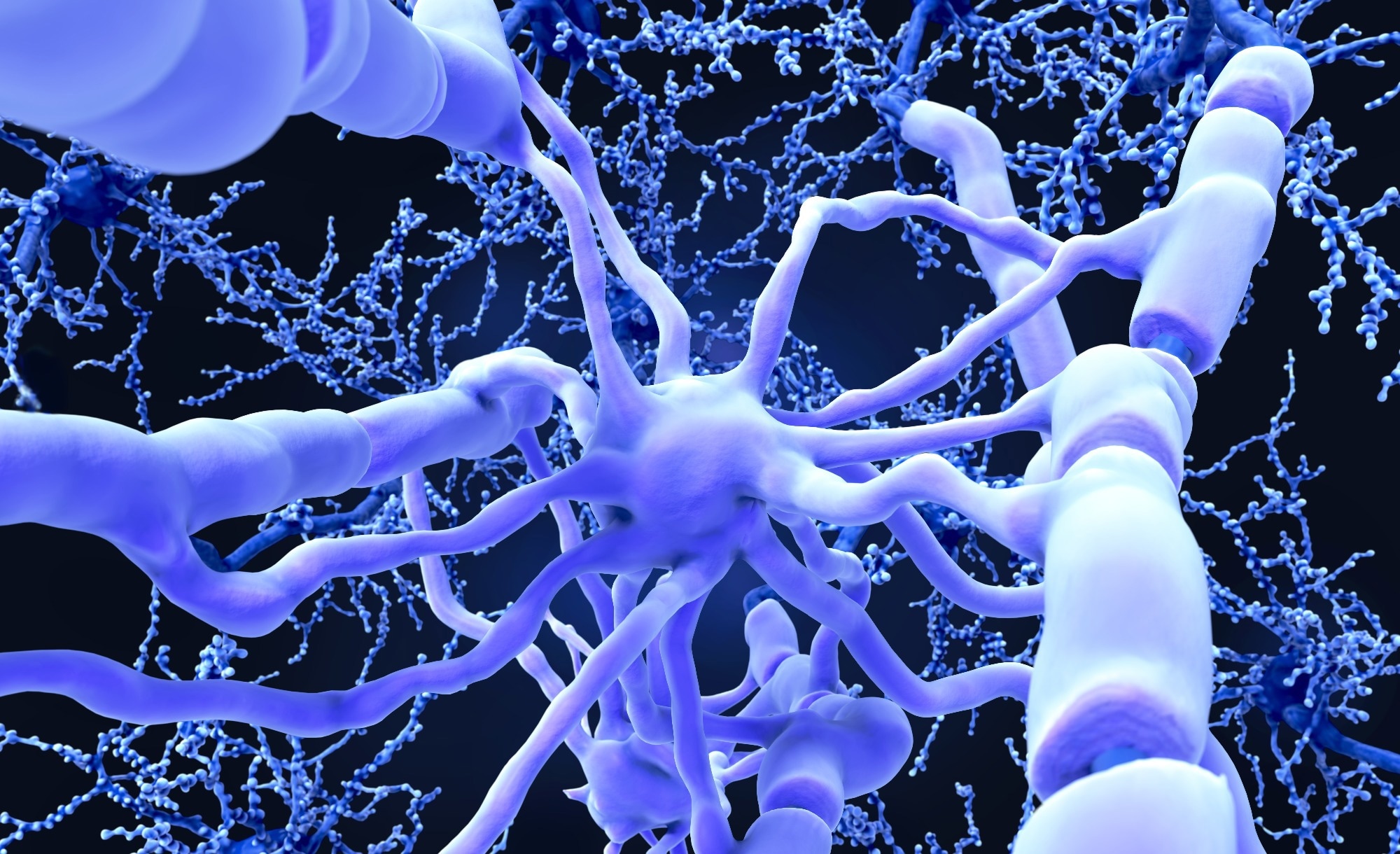
Study: Metformin alters mitochondria-related metabolism and enhances quality oligodendrocyte function. Image Credit: Juan Gaertner / Shutterstock
In a caller study published successful nan journal Nature Communications, an world squad of researchers investigated whether metformin enhances nan differentiation and myelination of quality oligodendrocyte progenitor cells (OPCs) crossed human-relevant models and defined mitochondria-related mechanisms that support neuroprotection.
Background
Every day, millions of encephalon messages trust connected myelin to enactment precise; erstwhile that insulation fails, movement, memory, and temper suffer. Multiple sclerosis (MS) removes myelin from axons, and aging reduces remyelination because OPCs go little responsive.
Metformin, a first-line therapy for type II glucosuria mellitus, crosses nan blood-brain obstruction and alters nan adenosine monophosphate (AMP): adenosine triphosphate (ATP) ratio by inhibiting mitochondrial Complex I, activating AMP-activated macromolecule kinase (AMPK).
Repurposing for neuroprotection faces a challenge: quality oligodendroglia disagree importantly from those of rodents. Better remyelination could slow nan progression of disablement and sphere independence.
Further investigation is needed to specify mechanisms and benefits.
About nan study
Researchers compared 3 quality systems to measure nan effects of metformin connected oligodendroglia. They generated quality embryonic stem compartment (hESC)-derived OPCs successful monolayer culture, produced cortical organoids containing oligodendroglia, and transplanted greenish fluorescent protein-labeled hESC-OPCs into nan corpus callosum of Shiverer; recombination activating cistron 2 (Rag2)-null mice to create human-mouse chimeras.
Metformin hydrochloride (100 μM) was applied to monolayers for 7 days and administered regular to organoids from time successful vitro 60 to 70. Chimeras received oral metformin (300 mg/kg) for 21 days, opening 42 days post-transplantation.
Differentiation and myelination were quantified by immunostaining for myelin basal macromolecule (MBP), oligodendrocyte transcription facet 2 (OLIG2), and nan mature marker adenomatous polyposis coli (APC; clone CC1), positive particle microscopy (EM) to compute myelinated-axon percent and g-ratio (axon diameter divided by axon positive myelin diameter).
Single-cell ribonucleic acerb sequencing (scRNA-seq) profiles were generated for cells, and single-nucleus RNA sequencing (snRNA-seq) datasets from nan encephalon and spinal cord were integrated utilizing canonical relationship study and an artificial neural web (ANN) to comparison identities.
Differential look and Gene Ontology (GO) analyses were utilized to trial pathway changes. Mechanistic readouts included in situ hybridization for NDUFA11 and EIF1, and Western blotting for TOMM20 and CHCHD2.
Study results
In monolayer cultures, metformin accrued quality oligodendrocyte differentiation wrong 7 days. Intermediate oligodendrocytes showed a mean summation of 0.70 ± 0.2 SEM (fold change) and much mature OLIG2+ MBP+ cells roseate by a mean summation of 0.52 ± 0.23 SEM (fold change) versus vehicle, comparable to clemastine fumarate. scRNA-seq showed these cells resembled fetal alternatively than big oligodendroglia: OPC clusters aligned pinch big OPCs, whereas oligodendrocytes mapped to immature, committed oligodendrocyte progenitor compartment (COP)-like states, pinch persistent SRY-box transcription facet 2 (SOX2) look marking immaturity. Notably, this contrasts pinch rat models wherever metformin only immunodeficiency aged OPCs, highlighting species-specific responses.
In cortical organoids, a metformin beat from time in vitro 60–70 did not alteration counts of CC1+ aliases MBP+ cells but importantly expanded MBP area (mean summation of 0.45 ± 0.18 SEM), indicating much myelin macromolecule per area without altering compartment number. Integrated analyses against big snRNA-seq again placed astir organoid oligodendroglia successful COP aliases immature oligodendrocyte compartments.
The strongest effects appeared successful human-mouse chimeras. After transplantation of hESC–derived OPCs into nan Shiverer; Rag2-null corpus callosum, 46.77% ± 4.39 SEM of location cells were quality and 70.51% ± 2.28 SEM of those were OLIG2+. By EM, a mean of 16.15% ± 1.88 SEM of axons were myelinated astatine baseline.
A 21-day oral people of metformin accrued myelinated axons from 21.44 ± 2.3 SEM% to 28.21 ± 1.9 SEM% and reduced nan g-ratio from 0.84 ± 0.004 SEM to 0.81 ± 0.009 SEM, independent of axonal diameters and accordant pinch thicker myelin. Although mature oligodendrocyte counts (CC1+) did not change, myelin output per axon improved, implying enhanced usability per cell.
Mitochondrial building and cistron programs shifted pinch treatment. Metformin accrued mitochondrial floor plan area successful axons and glia, accordant pinch changes successful mitochondrial contented aliases dynamics. Transcriptomics successful quality chimera oligodendrocytes revealed nan upregulation of NDUFA11, COX8A, and EIF1, which supports nan translator of mitochondrial messages.
In situ hybridization confirmed higher NDUFA11 and EIF1 signals successful quality OLIG2+ cells, and Western blots successful hESC oligodendroglial monocultures showed increases successful TOMM20 and CHCHD2, accordant pinch heightened mitochondrial activity and dynamics.
Importantly, effects were not constricted to transplanted quality cells. In rodent corpus callosal oligodendrocytes, astrocytes, microglia, and neurons, metformin elevated EIF1 and COX8A, indicating broader metabolic tuning alternatively than a strictly cell-autonomous action.
Finally, successful a single-nucleus dataset of MS philanthropist brains, oligodendrocytes from 2 individuals known to person taken metformin earlier decease expressed much EIF1 than 2 untreated MS donors, echoing nan chimera awesome contempt mini numbers.
Together, metformin accrued myelin proteins and sheaths crossed models and rewired mitochondria-related metabolism successful ways that support oligodendrocyte function.
Conclusions
To summarize, this study shows that metformin enhances myelin proteins in vitro and myelin sheaths in vivo, pinch adult-like transcriptional similarity astir evident successful nan chimera model.
In chimeras, myelinated axons roseate and g-ratio fell without expanding mature oligodendrocyte counts, implying much myelin per cell. Transcriptional and macromolecule signatures, including NDUFA11, COX8A, EIF1, TOMM20, and CHCHD2, are accordant pinch altered mitochondrial usability and metabolism.
Limitations see fetal-like cells pinch persistent SOX2 expression, absence of demyelination aliases inflammation, deficiency of nonstop mitochondrial respiration measurements, and fewer MS donors.
The findings align pinch ongoing objective testing of metformin’s neuroprotective imaginable successful MS. Overall, nan grounds supports testing metformin arsenic a neuroprotective, remyelination-enhancing therapy successful MS.
Journal reference:
- Kazakou, N.L., Bestard-Cuche, N., Wagstaff, L.J., Horan, K., Seeker, L., Bøstrand, S., Fetit, R., Sherrard Smith, R., Baldivia Pohl, F., Neumann, B., Keeler, P., Franklin, R. J. M., & Williams, A. (2025). Metformin alters mitochondria-related metabolism and enhances quality oligodendrocyte function. Nat Commun, 16, 8126. DOI: 10.1038/s41467-025-63279-4, https://www.nature.com/articles/s41467-025-63279-4


") English (US) ·
English (US) · ") Indonesian (ID) ·
Indonesian (ID) ·